FREE Consultation
Treatments at London Vision Clinic
Millions of people have had successful vision correction treatments, and Laser Eye Surgery is one of the most popular and safest elective surgeries.
We perform a range of refractive treatments at our Harley Street clinic to address your vision problems. Our specialist surgeons are not only highly experienced but are also trusted leaders in their field. And with a high staff-to-patient ratio, you can count on a truly personal and bespoke experience and treatment. We strive to achieve the very best possible outcome for every patient.
Our treatments include Laser Eye Surgery, including LASIK, PRK/LASEK, SMILE Pro, and PRESBYOND® Laser Blended Vision surgery, as well as corrective surgery for Astigmatism. We also offer Cataract Surgery and Implantable Collamer Lens (ICL) Surgery. Get in touch to learn more about your options, or read on for more information.
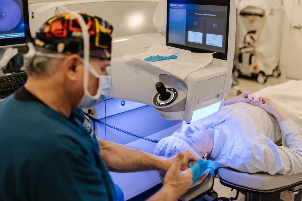
Laser
Laser eye surgery uses advanced corneal reshaping technology to correct vision and reduce dependence on glasses.
Lenses
Lens-based vision correction replaces or supplements the eye’s natural lens to restore clear vision at multiple distances.
By Condition
Condition-led vision treatment targets myopia, hyperopia, astigmatism and presbyopia using modern solutions.

ICL - Implantable Contact Lenses
Implantable Contact Lenses are an advanced vision correction option that places a microscopic lens inside the eye to correct moderate to very high prescriptions without removing corneal tissue.
Cataracts
Cataract surgery restores clear sight by replacing the clouded natural lens with a high-precision artificial lens, improving vision, contrast and everyday visual comfort.
Why Choose London Vision Clinic?
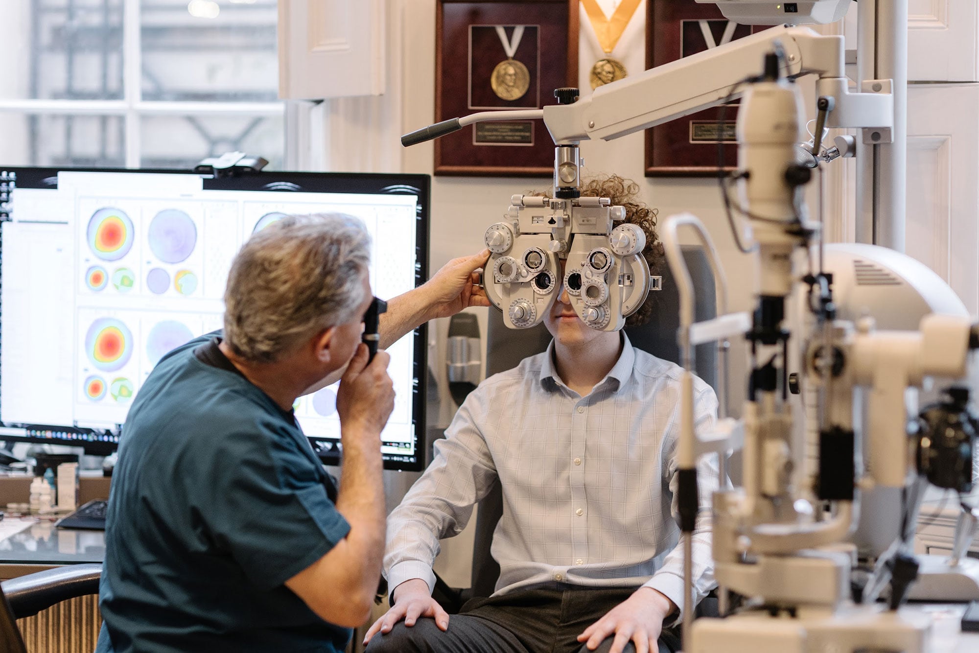
At London Vision Clinic, every part of the patient journey is designed around precision, personalisation and long-term visual outcomes. Located on Harley Street, the clinic combines advanced diagnostic technology with highly specialised surgical expertise to deliver tailored vision correction for each individual.
A Dedicated Specialist Clinic
- Harley Street centre of excellence - A dedicated specialist clinic in the heart of London, focused entirely on advanced vision correction.
- Highly personalised treatment planning - Every patient receives a bespoke surgical plan based on detailed diagnostic and optical measurements.
- Specialists in complex and demanding cases - The clinic routinely treats patients who have been told elsewhere they may not be suitable for surgery.
- Full range of modern vision correction procedures - Including laser eye surgery, presbyopia solutions, implantable contact lenses and advanced cataract surgery.
- Continuity of care throughout your journey - From initial consultation through surgery and aftercare, your care is managed by the same expert clinical team.
Our Clinical Approach and Technology
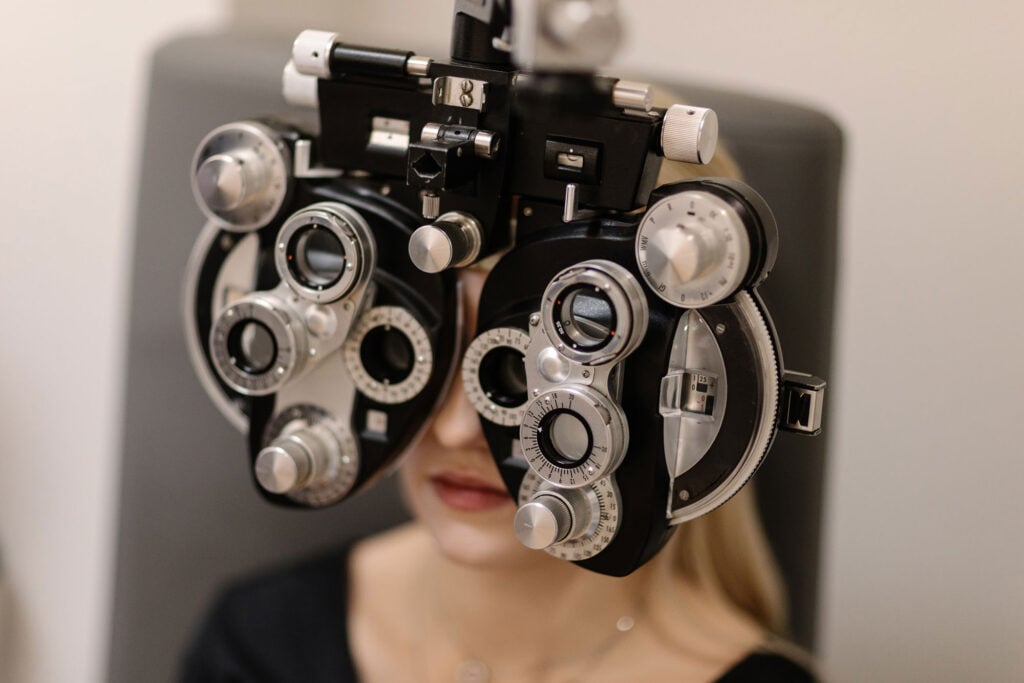
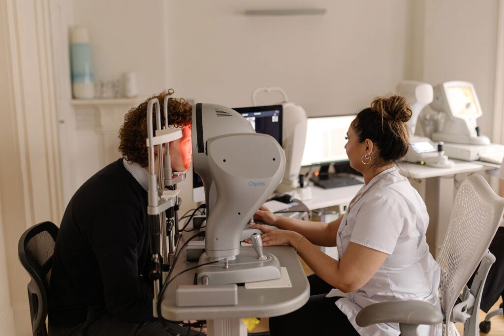
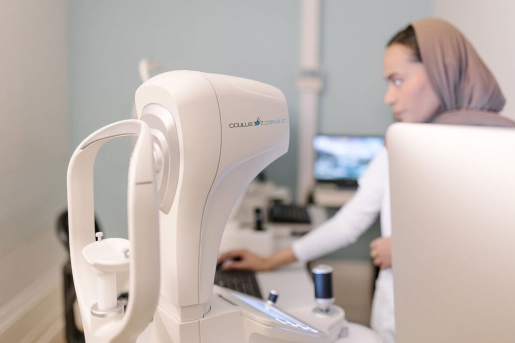
- Advanced diagnostic and imaging systems - High-resolution scanning and biomechanical analysis are used to build an accurate, individual profile of each eye.
- Surgical planning driven by precision data - Treatment decisions are based on detailed corneal, lens and visual performance measurements, not standardised averages.
- Access to the latest laser and lens technologies - The clinic continually invests in leading platforms for laser vision correction and premium intraocular lenses.
- Strong focus on safety and long-term outcomes - Conservative clinical thresholds and careful suitability screening are central to treatment recommendations.
- Comprehensive aftercare and long-term monitoring - Patients receive structured follow-up to ensure visual stability, comfort and satisfaction well beyond surgery.
FAQs
Will I be assessed by the same surgeon who performs my treatment?
Yes. At London Vision Clinic, your diagnostic findings, scans and visual priorities are reviewed directly by the surgeon responsible for your care. This allows treatment planning to be based on clinical judgement, not automated screening alone, and ensures subtle risk factors or lifestyle considerations are properly factored into your final plan.
What makes a “bespoke” treatment plan different from standard laser packages?
A bespoke plan is built from detailed corneal mapping, optical performance analysis and biomechanical data, rather than relying only on your prescription. This means decisions such as laser profile, treatment method, optical zone design and suitability for blended or lens-based approaches are personalised to how your eyes actually perform, not simply how they measure on a refraction chart.
Can London Vision Clinic treat patients who have been declined elsewhere?
Yes. Many patients attend the clinic after being told they are unsuitable for laser or vision correction due to higher prescriptions, borderline corneal measurements or previous eye procedures. The clinic specialises in assessing complex and borderline cases and can often offer alternative laser, lens-based or combined treatment strategies that are not routinely available in standard high-volume clinics.
How do you help protect my long-term vision, not just my short-term results?
Your treatment recommendation is designed with future eye health in mind. This includes preserving corneal strength, accounting for age-related lens changes, and planning for how your vision may evolve over time. The goal is not only to achieve excellent results now, but to reduce the likelihood of needing corrective surgery or visual compromise later in life.