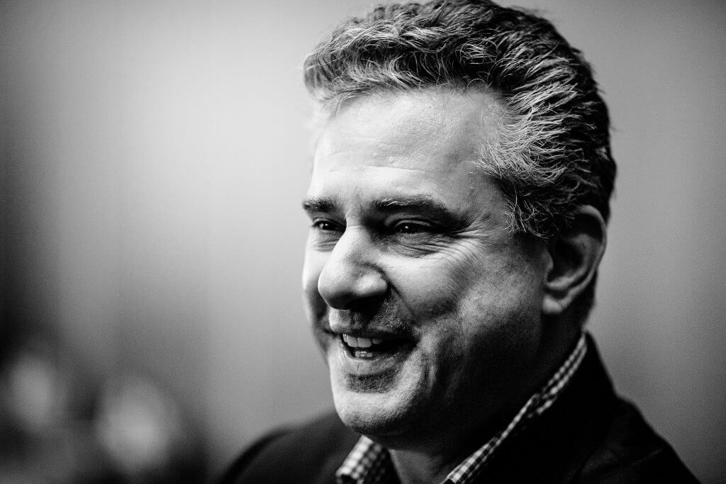
Sitting Down With… Dan Reinstein Medical Director, London Vision Clinic, London, UK.
Why medicine?
Being a doctor puts you in the extremely unique and privileged position to attend important moments in people’s lives – delivering a baby, being present when someone is told they have a fatal diagnosis, witnessing severe psychotic behaviour, or performing surgery. My career choice came down to choosing a specialty where I’d be using my academic strengths in math and physics, alongside my human strength of engaging with patients of all ages. I also wanted to become a surgeon, but needed a surgical specialty that would not impinge too heavily on evenings and weekends; I didn’t want to give up being a musician – something I started at age of four.
How do these two skills – music and medicine – complement each other?
Refractive surgery and jazz, to me, are analogous. Both require you to be able to make instantaneous decisions based on huge amounts of experience and expertise, as well as creativity and collaborative work – and of course, both give back extraordinary emotional satisfaction.
How did you end up taking the ‘long road’ to become a refractive surgeon?
Let’s define what we mean by long road: I extended my high school education by a year to sit the Cambridge entrance exam and was very fortunate to succeed in that, so I had a gap year before starting medical school. During this year I lived at my mother Erika Fabian’s house in Los Angeles, where I worked to convert her garage into a photographic studio for her and my stepfather, who was a National Geographic photographer. Simultaneously, I studied with a saxophonist, Alan Rowe, who was a graduate of the Berklee College of Music, which was one of the only jazz schools in the world at the time. At the end of this gap year, I spent four months as a professional jazz saxophonist in a top-40 entertainment band at a Club Med resort in Northern Morocco. I then did the first three years of medical school at Cambridge but having seen a crack in the door to the possibility of another year of music, I inserted a medium-sized crowbar and applied as much leverage as I could to get Cambridge to allow me to take a sabbatical to study at the renowned Berklee College of Music. I was fortunate enough to have tutors and a dean that gave me this leave of absence. A year at Berklee on a performance scholarship was possibly the hardest studying year of my entire educational career. But it was also extremely rewarding and resulted in my laying down the foundations for attaining virtuosity on the saxophone to last me a lifetime.
I returned to medical school for year four. Half way through I saw my new personal tutor, uroradiologist, David Rickards, and requested a second sabbatical to return to Berklee and complete their two-year diploma. I wasn’t expecting a yes, but Dr Rickards had himself taken time to sail around Africa as a trainee and he enthusiastically wrote to the dean, who in turn after a short interview, allowed the second year out of medicine. I spent the second year back in Boston and then finally returned to complete the last two years of medical school. I finished two years behind, graduating with final year prizes in ophthalmology and psychiatry. After my internships, I took six months of leave to study and formulate my postgraduate pathway training in ophthalmology. I figured that for jazz, I needed to be in New York and, as it happened, there were five residency training programs in the New York area, so it seemed like the obvious choice. I received advice that to make it possible as a foreign graduate, I would need to have done some research in the US to get into a residency program. So I applied to ultrasound pioneer, D Jackson Coleman, for a research fellowship at Cornell University Department of Ophthalmology bioacoustics lab, as it brought together my interest in ophthalmology, my strengths in computing, physics and math – all against the backdrop of New York City.
Towards the end of 1991, I became the first person to accurately measure the corneal epithelium in vivo in three dimensions with the 60 MHz very high-frequency ultrasound probes that had just arrived in the lab. I was literally euphoric to find that no one in the Department thought it was of any real clinical use – not until I received a call from Stephen Trokel at Columbia University the day he received some printouts of cornea ultrasound scans clearly resolving the epithelium. Within 48 hours we were scanning Phase II FDA PRK study subjects who were showing topographic ‘central islands’ and had no means of diagnosing the cause. This was to be the first of a long string of mysteries solved by what has become the Artemis Insight 100 Insight VHF digital ultrasound scanning technology. After the two year bioengineering research fellowship, I was admitted to the Icahn School of Medicine at Mount Sinai where I completed my ophthalmology residency program, while simultaneously retaining my position at Cornell to continue my ultrasound research in corneal imaging and biometry. After my residency, instead of going straight to fellowship training, I returned to the lab at Cornell (at less than half the salary of my contemporaries) to develop the very first ultrasound arc-scanner specifically for corneal and anterior segment imaging. Armed with this device, I found the perfect fellowship.
Hugo Sutton in Vancouver was a forward-thinking technology magnet who had been an earlier adopter of LASIK and had generated the expected number of learning curve complications. At the time, he was also one of the most experienced LASIK surgeons in North America. Working as a fellow under him, I was able to examine the library of misfortunes with my ultrasound device, gain 600 eyes LASIK experience as a fellow and go through a formal cornea fellowship training at UBC Vancouver, all within the span of 18 months. The next two years were spent as medical director of the very first corporate LASIK chain – LASIK Vision Canada – which grew to 30 clinics, 60 surgeons and performed 100,000 procedures in the last year before their business model collapsed due to poor financial controls. However, for the patients who were treated and the surgeons who participated in this, it was an extraordinary learning experience. Most of us were performing four or five thousand procedures a year with the leading technology of the time, under highly regimented operating standards. The proof of this was the largest peer-reviewed LASIK study, reporting a 50 percent sample of almost 100,000 procedures, published in the BJO.
Following the collapse of LASIK Vision, serendipitously I was contacted by Asclepion Meditec to move to Europe and work closely with the engineering team on the medical aspects of the excimer laser they were developing, which eventually became the MEL 80. The move to Europe resulted in my setting up my private practice, the London Vision Clinic, in 2002. So if I consider the pathway that I took from finishing school to starting my practice, by anyone’s standards that would count as a long road!
What is exciting you in the refractive surgery space right now?
There are three main areas. I feel like I’m surfing on the crest of a 50-foot wave in refractive surgery between SMILE and PRESBYOND® Laser Blended Vision. And, at the same time, I feel that I have reached a place in corneal refractive surgery safety where virtually every complication that may be generated can be returned to a good visual state. The other thing that is really exciting me is my increasing involvement in teaching – for example, publishing our textbook The Surgeon’s Guide to SMILE and running training courses in LASIK, SMILE and presbyopia correction. I get really excited when surgeons change their perspective on presbyopia, moving from clear lens exchange to PRESBYOND®, particularly given that nearly 20 of them have opted to have this on their own eyes, after years of following the crowd on dysfunctional lens syndrome.
You’ve had PRESBYOND® Laser Blended Vision LASIK surgery yourself – how did you feel?
It was an amazing experience on the one hand and an anti-climax on the other. Having treated thousands of patients myself with this technique, which I developed about 15 years ago, I didn’t expect many things to be a surprise – and they weren’t. I now see 20/12.5 at distance, J1 at near, J3 at intermediate, I have 40 seconds stereo vision uncorrected, and I am 100 percent spectacle independent at age of 56, having been a -0.75 D myope – or some might say plano-presbyope. But there was a moment that I wasn’t expecting. On the day of my procedure, I had been operating that morning. I was the last patient on the list. So I finished my last case and came back to the operating room – this time as the patient! When I lay on the bed waiting to start, it suddenly dawned on me that there was, in fact, a finite probability of something going horribly wrong with my vision. I then thought about the fact that my surgeon, Glenn Carp, had been my fellow for a year and a half and then worked with me for the next nine years, so I knew that there couldn’t be an intraoperative complication that he would not know how to handle perfectly. And then my mind went to postoperative complications. But I realized that he and I would be there to decide how to repair things. All of those thought processes took place in the 15 seconds it took for the bed to move into position under the cone of the VisuMax. I took a deep breath, felt very relaxed and enjoyed the show for the next 10 minutes!
The London Vision Clinic works closely with Carl Zeiss Meditec – what’s that like?
It’s like Christmas – you write a letter to Santa and you actually get some of those presents! I’ve really appreciated being able to contribute ideas for improving existing products or new products and features that I would like to use on my own patients. It’s a very privileged position to be in, at the interface between the surgical profession and the engineering profession.
Any notable career highs?
Being the first to measure and map the corneal epithelium. First realization that layered analysis of the cornea by ultrasound was the key to understand the complications of refractive surgery and therapeutic pathways to helping such patients. First to recognize the increased screening sensitivity and specificity of epithelial thickness mapping that can be added to topography and tomography to detect keratoconus at the earliest stages. First to understand the importance of trans-epithelial PTK digital subtraction pachymetry planning and epithelial power shifts. First machine algorithm for epithelial classification of pre-clinical and clinical keratoconus. First to develop spherical aberration control algorithms to increase depth of field and progress monovision to modified binocular vision and successfully treat all types of presbyopia in patients without cataract – clear lenses talked about as dysfunctional lens syndrome. First to realize the seemingly impossible task of bringing refractive surgery to one of the poorest countries in the world and developing a model whereby patients too poor to afford a pair of glasses could receive a permanent vision correction refractive surgical procedure in a cost-recovery model that is self-sustaining. The commercialization of the VHF digital ultrasound arc-scanner – from lab prototypes to Artemis Insight 100 1 and now the Insight 100. The completion of the SMILE textbook: The Surgeons’ Guide to SMILE.
What do you hope to achieve over the next 10 years?
I hope to develop software based on layered anatomical imaging of the cornea that will – as close to possible – enable any surgeon to correct any complication; to continue the quest to develop refractive surgery as a primary means of helping the poorest in the world suffering from refractive errors; and to play a lot more saxophone, hopefully kick-starting it with the release of my first record in 2019. In a wider sense, I also hope that refractive surgeons will learn to work as colleagues, not as competitors – to look after each other, our patient base, and the market so that refractive surgery becomes the standard for anyone suffering from refractive error disabilities.
With thanks to The Ophthalmologist Magazine.