A Brief Anatomy of the Eye
When trying to understand how Laser Eye Surgery treatments can effectively correct a whole host of vision impairments, it can be useful to learn a little bit more about the eye itself. As one of the most complex structures known to man, the eye remains something of a mystery to most people.
Despite using our eyes at every waking moment of the day, we rarely give a second thought to how this miracle actually works. On the other hand, modern advancements in science and technology mean that we now know more than ever before about these impressive organs.
The Structure of the Eye
The typical eye measures approximately just 24mm in diameter – and yet, they are the most complex organ in the body. Each eye is suspended in the socket with the help of a series of muscles and is protected by a thick layer of fatty tissue within the orbit.
Eyes are known as “paired structures” due to the collaboration of the eye itself and the surrounding muscle. These extra-ocular muscles allow us to move each eyeball in the same direction at the same time. This allows our brains to combine two slightly different images into a clear representation of the world around us.
This incredible ability, along with the help of our “binocular vision” (that’s the individual image from each eye), allows us to also determine depth and distance.
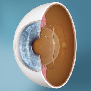
The eyeball itself is made up of three main components. The first component, known as the tunics, are the three layers of tissue that make up the wall of the eyeball. Second, are the optical elements that allow us to focus light; and the third is the neural components – the retina and the optical nerve.
For such as small organ, it really is amazing just how many working parts the eye has. So, let’s take a closer look at all these moving parts to help you better understand your own optical masterpieces.
Taking a peek at how your eye works
Let’s start with the most obvious question: How do our eyes actually allow us to see?
Our current understanding of vision is based on a model first devised in the 16th century. Felix Platter, a Swiss physician, first proposed that the eye functions as an optic, and the retina as a lens. Building on this early model, we now also know that light from an exterior light source enters through the cornea where a natural lens focuses it onto the retina – the light-sensitive membrane at the back of the eye.
From here, the retina sends signals along the optic nerve to the brain. The initial images that reach your brain are flat, 2D projections. Your brain works to overlay images from both eyes to form one seamless 3D image.
A slightly easier way to think about the workings of the eye is to compare it to a digital camera – after all, the theory behind cameras is the same as the function of the eye!
Light enters through the cornea as it enters through the lens of a camera. Tiny muscles then adjust the size of the pupil, much like the aperture of a camera, to control the amount of light entering the eye. This light is then reflected onto the retina – similar to how it is reflected onto camera film.
The retina contains millions of microscopic photoreceptor cells known as rods and cones. The rods are sensitive to light and dark, shapes, and movement while cones are sensitive to fine detail and colour. Together, they form detailed images that are transmitted to the visual cortex- the part of the brain that controls the sense of sight.
And just like that – you have your own ultra HD video feed of the world.
The Anatomy of the Eye
As we mentioned earlier, the eye has an incredible number of working parts – over two million to be more exact! These components work together to detect and process a staggering 35,000 pieces of information every hour.
You’ll be pleased to know that we aren’t going to attempt to cover every one of these components. We will, however, focus on a few of the main ones, starting with the cornea.
The Cornea
The cornea is the transparent dome-shaped structure on the front of your eye. It covers the iris and the pupil and allows light to enter the eye, making it incredibly important to our vision.
Not only does it transmit light into the eye, but the cornea also helps to focus light effectively towards the retina. In some cases, the cornea may not focus light effectively – this is known as ametropia refractive error. Laser Eye Surgery treats any refractive error by reshaping the tissue of the cornea to better focus light.
The Iris and Pupil
Immediately behind the cornea is the coloured ring membrane called the iris and the dark centre of the eye – the pupil.
In addition to determining the colour of our eyes, the iris contracts and expands to control the amount of light entering the eye. The pupil is the eye’s opening which works with the iris to regulate the amount of light that reaches the retina.
Situated behind the pupil is a colourless, transparent structure called the crystalline lens which also helps focus light.
The Ciliary Muscles
A collection of tiny muscles – known as the ciliary muscles – surround the eye’s lens. These muscles hold the crystalline lens in place but they also play an important role in vision.
When relaxed, the ciliary muscles pull and flatten the lens, allowing the eye to focus on objects that are far away. In contrast, when the muscles contract, the lens thickens and allows us to focus on objects up close.
Vitreous Humor
The largest component of the eye is known as the vitreous body – sometimes referred to simply as “the vitreous”. This is a transparent gelatinous mass that fills the space between the lens at the front of the eye and the retina at the back.
The vitreous connects many of the components of the eye. Light must pass through the vitreous humour before reaching the retina.
The Retina, Choroid and Sclera
The outer body of the eye is made up of three layers – the innermost layer being the retina. This vital component contains over 130 million receptors which allow it to process accurate images.
The outer layer extends nearly all the way around the eye – it is called the sclera. The sclera is what provides the eyeball with the majority of its white colour.
Finally, the middle layer, called the choroid, contains the blood vessels that supply the eye with oxygen and nutrients and help to remove waste products.
The Optic Nerve
The last of the main components we are going to look at is the optic nerve. This nerve is part of the central nervous system and contains over a million nerve fibres. The vast majority of these fibres play a role in conveying information related to central vision.
The optic nerve starts at the back of the eye as the optic disc – a structure approximately 1.5mm in diameter. As it leaves the back of the eye, the optic nerve passes through the eye socket and the bony optic canal until it reaches the underside of the front of the brain. It is responsible for carrying the information from the retina to the brain where our image of the world is finalised.
And there you have it! From photons of light, our eye is able to create a crisp image – all in less than a second. Your vision truly is one of life’s great miracles.
If you’d like to find out more about your eyes and vision, why not Book a Consultation and receive a comprehensive eye test? Get in touch today and speak with one of our friendly and knowledgeable clinic coordinators.