There are few things in the natural universe more complex than the eye. This vital organ may be small but it works using an abundance of processes and components – all of which must be running smoothly – to give us a clear image of the world around us. In this role, our eyes have become one of the greatest assets we have – an invaluable part of our bodies and lives. So, how does this incredible organ work? And what happens when things aren’t working quite as they should?
In this article, we’ll be answering these questions in more detail. We’ll touch upon the mechanisms of this incredible organ and take a closer look at the components that make up nature’s own camera.
Your Eye is Like a Camera
You may have heard this comparison made before – and with good reason. In fact, the design of the first camera was based on our understanding of how the eye works. This concept still stands today, making the camera a perfect metaphor for our most valuable organs.
Our eyes measure, on average, less than 2.5 cm. Yet, despite their small size, they are – aside from the brain – the most complex organ in our bodies. Your eye consists of a number of vital components:
- The pupil: a self-adjusting opening in the front of the eye;
- A lens system: this includes a transparent covering (the cornea), which controls most of the eye’s focusing power, and a spherical lens inside the eye (behind the iris);
- The retina: a light-sensitive layer of tissue at the back of the eye that sends signals to the brain;
- Various muscles: these control the dilation and contraction of the pupil, the shape of the lens, and the movements of the eye.
Much like the different components of a camera, these elements all work in unison to send to your brain a representation of the world around you. But how does this actually work?
The Process of Vision
Light plays an integral part in how we see things. It bounces off all the objects around us and this reflected light enters our eyes through the pupil. On its way, the cornea refracts this light in order to place it effectively on the retina – which can be compared to the film in a camera. The retina contains many sensory cells called ‘rods’ and ‘cones’ which change the photons of light into electrical signals.
- Nerves attached to the eye then transmit these electrical signals to the brain, where they are interpreted as an image.
- This might sound like magic, but in order for our brain to create a clear image of what we see, four crucial things must happen:
- The image must be ‘reduced’ to fit onto the retina;
- The scattered light must be effectively focused onto the surface of the retina;
- The image must curve to match the natural curve of the retina;
- The brain must interpret the image as vision.
A lot of work goes into meeting these four requirements. To achieve these steps, intraocular muscles must work to contract and relax the shape of the lens system to keep light correctly focused on the retina. These muscles are controlled by your nervous system. Sometimes, errors can occur that cause these muscles to work ineffectively. This can result in visual impairment, such as in presbyopia.
Most vision problems occur when the eye is unable to effectively focus images onto the retina.
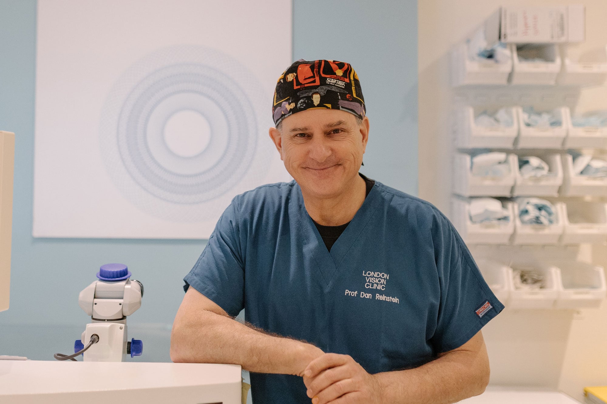
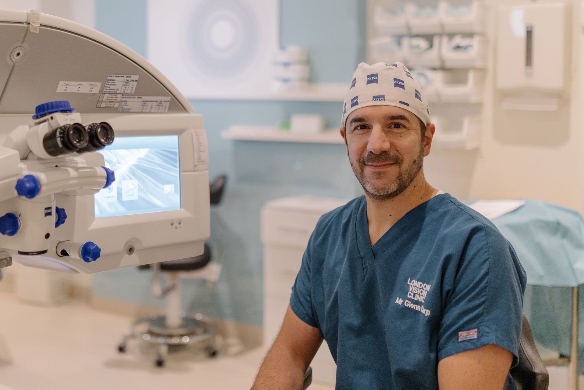
Many common refractive errors – including long-sightedness (hyperopia), short-sightedness (myopia), ageing eyes (presbyopia), and astigmatism – are caused by an irregularly shaped cornea, the length of the eye, and/or the stretchiness, or ‘elasticity’, of the lens. Luckily, in the vast majority of cases, these errors can be corrected with the help of Laser Eye Surgery.
If you are interested in learning more about how Laser Eye Surgery could treat your refractive error, get in touch with one of our friendly clinic coordinators. Alternatively, Book a Consultation online, today.
[guide_form]